Retina Vascular Occlusions

INTRAVITREAL ANTIVEGF/OZURDEX INJECTIONS
MICROPULSE SUBLIMINAL LASER PHOTOSTIMULATION
PRP GROWTH FACTOR INJECTIONS
MAGNOVISION THERAPY

Retinal veins may occlude in patients with hypertension, diabetes, cardiovascular disease or glaucosis. This occlusion may occur in only one branch of the vein as it also may occur in the form of retinal root vein occlusion in the exit of optical nerve.

Vascular pressure rises behind the occlusion. Bleeding and edema occur in the retina. Blurred and hazy vision occurs, coinciding with the area of bleeding and edema.

In fundus fluorescent angiography, which is to be performed after the hemorrhages are partially removed, the presence of ischemia, that is, the nutritional status of the retina, is investigated. In the absence of ischemia, the results are usually better.
If there is ischemia in retinal vein root occlusion, new blood vessels are formed within 3 months, depending on these, intraocular bleeding and a painful and difficult-to-treat glaucoma called 100-day glaucoma (neovascular glaucoma) may develop.

Intraocular injection treatments, micropulse or grid/panretinal thermal laser treatments are applied if necessary to prevent new vessel formations and/or macular edema.
If ischemic damage to the retina is extensive, magnovision therapy and extraocular growth factor injection support may be required.
The underlying cause should be investigated; hypertension, diabetes, glaucoma must be kept under control.

Retinal artery (arterial) occlusion

Occlusion develops in the artery that feeds the retina, as a result of the formation of fatty plaques or a clot from the carotid.
If the occlusion occurs in the part of the optic nerve, retinal main artery occlusion occurs. Painless and sudden loss of vision occurs.
Its treatment is very urgent. If the clot cannot be dissolved or moved further within 4 hours, permanent vision loss develops.
In the treatment, intraocular pressure is lowered and subcutaneous low dose heparin is started. Vasodilators are administered. If possible, hyperbaric oxygen therapy is started immediately. Hyperbaric oxygen both provides dilation of retinal vessels and meets the oxygen requirement of the retina.
Magnovision therapy to be applied as soon as possible and extraoccular growth factor injection support contributes to the repair of retinal damage.

If the arterial occlusion occurs in only one branch, retinal ischemia, that is, the damage, develops only in the retinal region supplied by that branch.

Macular Degeneration (Senile/Age-Related Macular Degeneration)

INTRAVITREAL ANTIVEGF INJECTIONS
PRP GROWTH FACTOR INJECTIONS
MAGNOVISION THERAPY

The eyeball consists of refractive settings that focus light on the retina from front to back. The cornea in front, and the lens behind it focuses light on the macula, the most sensitive area of the retina.
The eyeball consists of 3 layers from the inside out: the retina is the innermost, the vascular layer in the middle, and the white of the eye, which gives the eyeball its rigidity, is the outermost.
Objects can be seen when the rays coming from the objects focus on the retina. There are cells in the retina that convert light energy into electrical energy and transmit it to the brain.
The region called the macula or yellow spot is the region where the light is fully focused and the sharpest vision occurs. The image is transmitted to the brain most clearly by the cone cells in this area. 90% of visual acuity is formed in the yellow spot and 10% is formed in the surrounding retina and then transmitted to the brain.

The yellow spot is the retinal region where lifelong high light-chemical and metabolic reactions occur. With increasing age and genetic predisposition, metabolic residues cannot be cleared from this region after the age of 50 and begin to accumulate. These yellow dots that accumulate in the retinal layers are called drusen.

In electron microspic sections, metabolic wastes are seen to accumulate under the retinal pigment epithelial cells in the form of fat deposits called drusen.
These accumulated fats prevent the retinal pigment epithelium from receiving oxygen and nutrients from the large choroidal vessels. The cells above the fat deposits die. This condition is dry macular degeneration.
The pigment epithelium has stem cell characteristics and these cells secrete growth factors for the viability of photoreceptor cells. When these cells die, the photoreceptor cells above them also go into sleep mode. In order for these cells to continue their viability, new glomerulus are formed. However, since these glomerulus are fragile and thin, they cause bleeding and leakage from the vessels, leading to the damaging of photoreceptor cells.

This condition is wet macular degeneration.

The stage in which fat cells called drusen accumulate and pigment epithelial cells enter sleep mode is called early-stage dry senile macular degeneration (SMD).
Diagnosis is very important during this period. Very serious measures should be taken to stop the progression of the disease, with these, the course of the disease can be slowed.
If the disease progresses further, the pigment epithelial cells begin to die. In this case, central vision is reduced. This stage is called atrophic, that is, dry SMD.

If new vessel formations occur in the area where the pigment epithelial cells die, this stage is called wet SMD. This is the most critical stage, and it causes severe vision loss.
At this stage, medication injections called anti-VEGF are administered into the eye. With this, it is aimed to shrink/close the new vessels that are suitable for bleeding and to reduce/remove the edema in the macula. Otherwise, significant hemorrhage may develop under the macula and into the vitreous.
Approximately 8 intravitreal injections are required in the first year, and these injections are often effective. However, there are cases that are resistant to injection. In this case, the drugs used for injection are changed or combined.

Again, if there is no response to the injections, “photodynamic laser therapy” should be considered, in which a special drug is injected into the arm’s vein and at the end a special laser is applied that only closes the damaged new vessel formations.
If unwanted glomerulus cannot be controlled, excessive bleeding occurs in the eye.
When bleeding recedes, it is seen that all retinal layers in the macula are damaged, forming scar tissue and atrophy (disciform scar stage). As a result, central vision is severely affected and the patient sees only with their peripheral retina.

Symptoms, complaints and management of macular degeneration
It is very important for early diagnosis that everyone over the age of 50, especially with a family history of macular degeneration, should have an eyeball check at least once a year.
The patient may not have any complaints in the early stage, that is, in the stage where the fats called drusen start to accumulate in the yellow spot. At this stage, for smokers to quit smoking, controlling hypertension and cholesterol and fighting obesity severely slow down the rate of progression of the disease.
Again, in the early stage, the intake of nutritional supplements rich in antioxidant minerals and vitamins and the intake of vitamin complexes will slow down the course of the disease. B12-b6-b1- folic acid and omega-3 are especially necessary for the vital activities of neuronal cells, while vitamin a-lutein-carotene is necessary for the synthesis of pigments used in photoreceptors to perform their vision function.
In particular, carbohydrate and sugar consumption should be avoided.

Mediterranean diet

  • Grilled salmon: 2 days a week (all fatty fish are allowed, provided that they are grilled-fried fish has no benefits.)
  • Plenty of green salads + grated carrots
  • Soft-boiled eggs: 3 days a week (boiled egg with apricot consistency)
  • Raisins with black seeds (fresh when in season) : 1 handful 1 day a week
  • Raw almonds: 1 handful 1 day a week
  • Walnuts: 1 handful 1 day a week
  • Kefir: 1 glass 2 days a week
  • 30 minutes of walking outdoors every day
  • Absolutely no smoking
  • Abstinence from alcohol

should slow the rate of progression of the disease considerably.

Protective glasses:
Filtering of blue-violet light that are at a wavelength of 415-455 nm reduces the photoreceptor death rate by 25%.
500 nm orange lenses are recommended for patients suffering from excessive glare where the initial cataract or cone cells are also affected.
In the early stage, amsler grid test is used for self-examination and follow-up of patients at home.

After wearing reading goggles in a well-lighted room, look at the squared paper that your doctor will give you first by covering one eye with your palm and then doing the same for the other eye few times a week.
If there are refractions in the lines or inability to see the middle point in the center, the disease has passed from the early stage to the advanced stage. Consult your ophthalmologist as soon as possible.
If the diagnosis is of atrophic type, the retinal pigment epithelium is usually in sleep mode at this stage. At this stage, the pigment epithelium is awakened with a micropulse-sub-threshold laser. With the application of this non-burning laser, both growth factor release and biophotomodulation are provided, and the pump function of the pigment epithelium is stimulated and the drusen clearing is accelerated. At this stage, together with micropulse laser, growth factor injections can also be applied to the outside of the eyeball. Magnovision support is applied if necessary.

If straight lines such as door edges or flagpoles appear refracted or loss of central vision has begun, it means that the wet stage has begun.
New vessel formations in the wet stage; it should be dried with intraocular injection treatments and, if necessary, photodynamic laser treatment. Otherwise, very serious vision loss occurs at this stage.
If permanent loss of central vision occurs; low vision rehabilitation devices such as telescopic glasses, light magnifiers, cctv camera systems will especially facilitate reading.
Implantable miniature telescope surgeries may be a good option in appropriate cases for patients who have lost their central vision in both eyes due to macular degeneration.
At the end of the cataract operation, these special lenses are placed inside the eyes and these lenses focus the image on the yellow spot.
With a prismatic effect, these telescopes magnify and shift the image to an intact retinal area other than the damaged yellow spot.

Diseases Of Optical Nerve (Visual Nerve) And Optic Tracts (Optic Neuropathy)

PRP GROWTH FACTOR INJECTIONS
MAGNOVISION THERAPY
PHOTOBIOMODULATION THERAPY

Vision function begins when rays from objects are focused on the retina. Light energy is converted into electrical energy in the retina. These electrical impulses are transmitted to the visual center in the brain by passing through the optic nerve, optic chiasm and optic tract called visual pathways. Nerve fibers emerging from the retina merge to form the optic nerve.

The optic nerve can be damaged for various reasons and vision or field of vision loss may occur.
Causes of optic nerve disease:

Compression of the optic nerve: due to thyroid (goiter) diseases
Infection of the optic nerve: most commonly due to otitis media and sinusitis
Ischemia of the optic nerve, i.e. occlusion of the vessels feeding the optic nerve
non-arteritic (diabetes, hypertension is the most common cause)
arteritic (due to arterial inflammation: this is a very severe condition,
Blindness in both eyes may occur if treatment is not started immediately.
Traumatic cut or injury of the optic nerve
Damage to the optic nerve sheath in diseases progressing with loss of nerve sheath called multiple sclerosis.
Toxic effects on the optic nerve due to alcohol (methyl alcohol), tuberculosis drugs such as ethambutol-isoniazid, damage due to radiation therapy
Damage to the optic nerve as a result of some systemic diseases such as lupus-Behçet’s
Optic nerve atrophy as a result of some genetic-familial diseases such as Leber’s disease
Optic nerve damage due to eye pressure, i.e. glaucoma
Apart from the eye, damage to the optic nerve and visual pathways may occur due to neurological diseases:

Papilledema and related optic atrophy as a result of increased intracranial pressure (ICP)
Pressure on optic chiasm by pituitary gland adenoma or tumors
Occipital lobe and visual cortex problems
Migraine
Bubbling in brain vessels (aneurysms)
Brain tumors
Infections such as meningitis-encephalitis

Depending on the location of the damage in the optic nerve and visual pathways, various types of losses occur in the field of vision.

In optic nerve diseases, patient sees the objects hazy, gauzy and pale; senses field of vision losses.
The light reflex is weakened on the affected side and contrast sensitivity, color vision and visual field are impaired.
Treatment: Depending on the underlying cause, it is done by the relevant branch of medicine.

In optic neuritis due to multiple sclerosis, there is damage to the myelin sheath that supports nerve conduction.

In ischemic optic neuropathy, the treatment modality is determined after the arteritic/non-arteritic distinction is distinguished by a high sedimentation rate. If it is arteritic, high dose and long-term cortisone is required to protect the other eye and if it is non-arteritic (sugar-blood pressure control), depending on the cause, blood thinners are applied together with cortisone. Magnovision and growth factor support may be required to increase optic nerve blood flow.
In trauma-related optic nerve diseases, magnovision and growth factor treatments with cortisone are urgently applied.

The cause of optic nerve disease can be understood with other examinations, especially MRI.

Optic atrophy (weakening, drying of the nerve) may occur as a result of various optic nerve diseases. In this case, new visual pathways can be activated by stimulating the weakened visual nerves with magnovision/okuvision.

If the eyes and visual pathways are completely damaged, it seems that all blindness can be cured in the near future by applying stimulating chips, that is, cortical prostheses, to the visual center in the brain. Experimental studies on this subject are ongoing, and there is no routine and widespread application yet.

Retinitis Pigmentosa (RP) Night Blindness

PRP GROWTH FACTOR INJECTIONS
MAGNOVISION THERAPY
PHOTOBIOMODULATION THERAPY
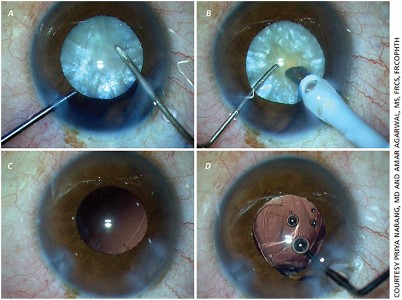
ARGUS II RETINAL PROSTHESIS OPERATION

The eyeball consists of refractive settings that focus light on the retina from front to back. The cornea is at the front and the lens behind it focuses the light on the retina.

The eyeball consists of 3 layers from the inside out. The innermost retina is the vascular layer in the middle, and the outermost is the white of the eye that gives the eyeball its rigidity.

Objects can be seen when the rays coming from the objects focus on the retina. There are cells in the retina that convert light energy into electrical energy and transmit it to the brain. These are called photoreceptors. Photoreceptors consist of two groups:

  1. Cone cells: images formed in the center and in a bright environment,
  2. Rod cells: processes images formed in the surrounding and dark environment.

In other words, they convert the rays coming from the objects into electrical energy.

Electron microscope view of the section taken from the retina. Just above the lowest pigment epithelium are photoreceptor cells consisting of rods and cones.

Retinitis pigmentosa begins with a mutation, or defect, in the genes of photoreceptor cells.

250 different genetic mutations lead to 50 different types of this disease. For this reason, clinical symptoms and progression rate are different in each patient. It is usually familial due to consanguineous marriage, but it can also occur non-familial.

Genetic mutation leads to premature aging and progressive death of photoreceptor cells. As age increases, cell death accelerates.

Growth factors enable the conversion of glucose in the blood to ATP-GTP, which is the cell fuel. In the absence of growth factors, photoreceptor cells slow down their metabolic activities due to energy deprivation, go into sleep mode, and if the process continues, cell death occurs progressively.

The growth factors that keep the photoreceptors alive are not secreted by the pigment epithelium and Müller cells, which show stem cell characteristics. Photoreceptors go into sleep mode for a long time before slowly dying. At this stage, the cells are alive but do not participate in the function of vision.

Which cells go into sleep mode or die first, symptoms begin accordingly. For example, if the rod cells start to die first, the first symptom will be reduced night vision.

In some genetic types of the disease, cone cells are affected before rod cells are affected. The cone cells first go into sleep mode and then die.

This condition is seen in stargarth disease, reverse rp, or cone dystrophy.

Here, the first symptom is excessive glare in the light, and better vision in dim environments and at night. As the disease progresses, central vision is lost and peripheral vision is preserved.

Retinitis Pigmentosa Treatment:

1) Stem Cell and Gene Therapy :

Its studies are still in progress and the target group is the patient group who has not completely lost their sight and has living cells in sleep mode. The rationale of stem cell therapy is to inject cells capable of secreting growth factor between 6 months and 2 years, into or around the eye, and repeat this process every 6 months or 2 years.

In gene therapy, the faulty gene in photoreceptors that have not lost their vitality is detected, the corrected gene is loaded into viruses and injected into the eye. The aim is again to stop the death of living photoreceptors.

2) Early diagnosis:

The earlier the diagnosis is made, the death of the remaining cells may be slowed down as much as possible without further loss of photoreceptors, may be stopped, or even cells in sleep mode may be awakened.

3) Growth factor injections

Ngf, bdnf, bfgf, IL6 are growth factors necessary for photoreceptors to maintain their vitality and functions.

First loading and then re-injection at certain intervals may slow down or stop the course of the disease. In some cases, depending on the presence or number of cells in sleep mode, visual field enlargement and visual acuity may be increased.

Enlargement of the visual field after the 3rd injection

4) Okuvision & Magnovision

Electromagnetic stimulation of the retina and the visual center in the brain not only increases the secretion of the necessary growth factors in the nervous system, but also allows the formation of new visual pathways called neuronal plasticity and the reorganization of visual function.

The awakening of the photoreceptors in the sleep mode and the enlargement of the visual field in the patient who underwent growth factor injection together with magnovision are shown in the figure below.

5) Treatment of complications

Cataract develops frequently in rp patients. In advanced stages, retinal vessels become thinner and oxygen reaching the intraocular tissues and essential nutrients decrease. This accelerates the development of cataracts.

Cataract operation in rp patients can accelerate cell death in the retina and cause excessive increase in macular edema. For this reason, protecting the retina with growth factors before cataract operation will minimize the possible risks.

Macular edema is another common complication in rp patients. Edema at a certain level does not require treatment as it meets the growth factor requirement for photoreceptors. However, edema that impairs visual acuity and becomes cystoid requires treatment. If it cannot be controlled with eye drops, edema should be controlled with intraocular injections.

6) Safety glasses

Filtering blue-violet light at a wavelength of 415-455 nm reduces the photoreceptor death rate by 25%. It is especially recommended for computer users.

500 nm orange lenses are recommended for rp patients with initial cataract or excessive glare in the light where cone cells are also affected.

7) Low vision rehabilitation tools:

In patients whose cone cells are affected and peripheral vision is good, telescopic glasses, magnifiers with light, or magnifiers with CCTV cameras respond particularly well to the reading needs of the patients.

8) Intraocular telescopic lens implantation:

Prismatic telescopic intraocular lenses, which shift the image out of the dead area, give satisfactory results in suitable patients in patients whose cone cells are affected and peripheral vision is good.

9) Nutrition-healthy life:

It is possible to slow down the death rate of photoreceptors by consuming foods high in antioxidant vitamins and minerals and food supplements. B12-b6-b1-folic acid and omega-3 are especially necessary for the vital activities of neuronal cells, while vitamin a-lutein-carotene is necessary for the synthesis of pigments necessary for the visual function of photoreceptors.

Mediterranean Type Nutrition

  • Grilled Salmon: 2 days a week (all fatty fish – on the condition that they are grilled or baked)-(fried fish has no benefits.)
  • Plenty of green salad + grated carrots
  • Soft-boiled eggs: 3 days a week (boiled eggs in apricot consistency)
  • Raisins with black seeds or fresh black grapes in season: once a week, handful
  • Raw almonds: once a week, handful
  • Walnut: once a week, handful
  • Kefir: 2 days a week, 1 glass

30 minutes of walking outdoors every day

Absolutely no smoking

Abstinence from alcohol

It will greatly slow down the death rate of photoreceptors.

10) For patients whose photoreceptors have completely lost their vitality:

Virtual vision and rehabilitation with chips to perform the function of photoreceptors.

The image is perceived by a camera – converted into electrical energy and transmitted to the brain by electrodes. For this,

  • brainport v200
  • argus 2 bionic eyes
  • cortical prosthesis

The most appropriate method is applied to the appropriate patient.

Retinal Detachment

PATTERN LASER
ENDOSCOPIC 25 PLUS PARS PLANA VITRECTOMY SURGERY

A transparent and viscoelastic substance called vitreous fills in the eyeball. This substance gives the eye a spherical shape and brings it elasticity. The vitreous gel exhibits tight attachments to the retinal layer in some areas.

As a result of advanced age, high myopia, some impacts to the eye or head trauma, the vitreous gel begins to liquefy and detach from the retina. During this separation, tears or hemorrhages may occur where the vitreous attaches to the retina. As a result of genetic predisposition, thinned degenerated areas sensitive to tearing may be present in the retina (lattice, tuft). Retinal tears can also form as a result of some traumas, cataract surgery or other eye surgeries.

Liquefied vitreous passing through these tears in the retina causes the retinal layers to detach from each other. Detachment and/or fluctuations together with degeneration of the vitreous in the retina, which is a nervous tissue, causes symptoms such as flashes of light, flying flies or spider webs. A curtain over the eye is mentioned when the retina, under which the fluid accumulates, is detached from the eye wall. As this detachment affects the macula, vision decreases to the sense of light.

If retinal detachment is not treated almost immediately, the fluid may advance through the retinal layers, causing detachment of the entire retina and permanent vision loss.

Retinal detachment may directly give symptoms such as rapid closure of vision without flying flies, flashes of light, loss in visual field, or any of these.

Flying flies, flashes of light, and the feeling of floaters may sometimes occur only in the presence of retinal tears without detachment. At this stage, the development of detachment can be prevented by attaching the periphery of the tear with a barrage laser without any fluid entering between the tear.

If retinal detachment has developed due to detachment of the retina from the eye wall; depending on the condition of the eye and the decision of the ophthalmologist, either standard retinal detachment surgery or vitreoretinal surgery (pars plana vitrectomy) surgery is decided. These surgeries are usually performed with general anesthesia. During the surgery, it is decided by the physician to perform intraocular tamponade with medical silicone oil or special medical gases. In the postoperative period, it may be necessary to maintain a certain position and lie down in that way for 7-10 days, which will be determined by the physician.

Repair of retinal detachment and tear with pars plana vitrectomy surgery

Until the tear can attach completely, sterile gas or air is injected into the eye and pressure is applied on the tear. After the operation, the patient is provided with a head and lying position so that the buffer gas can come over the tear.

In some cases, it may be necessary to apply compression to the (sclera with special materials for complete closure of the tear.
Retinal detachment most commonly occurs as a tear retinal detachment.
Tractional retinal detachment may also develop as a result of fibrotic bands pulling the retina from the inside, after retinal vascular diseases or some trauma-intraocular foreign bodies in diabetic retinopathy.
Exudative retinal detachments may develop in malignant and some benign intraocular tumors, intraocular inflammations called uveitis, eclampsia during pregnancy, renal failure and high systemic hypertension.
Anatomical success rate is very high after retinal detachment surgeries. The earlier the intervention, the higher the chance of success.
If the retina is damaged, growth factor injections outside the eyeball or monovision support can be applied after the surgery.

Cataracts

PHACOEMULSIFICATION
IOL

Behind the colored part of the eye iris, there is the lens of the eye.
The lens is glass-like transparent and it allows the rays from objects to focus on the sensitive point of the retina called the macula.
It is called cataract when the lens loses its transparency over time and becomes obscure.

Lens with cataract seen through the clear lens pupilla

Cataract clear lens

Causes of cataract:

  • Cataract most commonly occurs after the age of 60, due to tissue aging.
  • It can be seen at earlier ages in conditions such as diabetes, working in hot environments, trauma, long-term use of cortisone.
  • Sometimes it can occur at birth or in early childhood. Congenital cataracts may develop due to genetics or some infections that the mother had during her pregnancy and some drugs used.

Patients with cataracts complain of veiled-foggy vision, seeing as if looking from behind a waterfall, double vision with one eye, glare in the light, dispersion of lights at night, and complete closure of vision.

Treatment of cataract:
The only treatment for cataract is surgery. The clouded intraocular lens is cleaned by liquefying with the phacoemulsification process.
The natural lens has a refractive optical property of +22 on average. Objects can only be focused in the macula with this optical refraction. After the cataract lens is cleaned, an artificial and transparent intraocular lens is placed into the eye to provide this refraction. If the artificial lens is not placed, the patient cannot focus the image and sees blurred.

Cataract surgeries are surgeries that are performed under a microscope and require advanced technology.
Artificial intraocular lenses can be unifocal (far or near) or multifocal for far and near. However, single-focal lenses should be preferred in patients with retinal diseases along with cataracts. Because the contrast sensitivity of multifocal lenses is low. It also makes it difficult for the retina to appear clear and to be treated.

In patients with macular degeneration or macular damage, vision can be improved by placing miniature telescopic lenses in the eye during cataract surgery.

Cataract surgery is an operation that is mostly performed under local anesthesia.

Cataract surgeries can also be performed in the same session with vitrectomy surgery for the treatment of retinal diseases. In this type of surgeries, general anesthesia is preferred since the operation will take a long time.

Glaucoma (Eye Pressure)

LASER TRABECUOLOPLASTY
LASER CYCLOPHOTOCOAGULATION
LASER IRIDOTOMY – PRP GROWTH FACTOR INJECTIONS
MAGNOVISION THERAPY

Eye pressure i.e. glaucoma is a progressive disease that can cause permanent vision loss if left untreated.

In glaucoma, there is an increase in intraocular pressure to a level that damages the optic nerve, which is necessary for vision, as a result of the inability to drain the intraocular fluid that is constantly made for the nourishment of the intraocular tissues.

It often starts over the age of 40 and usually does not show any serious symptoms until permanent vision damage occurs.

For early diagnosis, it is extremely important for individuals with a family history of glaucoma to go to an ophthalmologist regularly from the age of 35-40 and have their eye pressure measured.

Glaucoma can generally only be noticed by the patient in the advanced period. Early diagnosis is very important, as the damage that occurs after vision loss is irreversible.

High intraocular pressure detected during a normal eye examination may be the first sign of the disease.

Ophthalmoscopic view of the normal fundus.

In uncontrolled glaucoma, extreme damage to the optic nerve and progressive field of vision loss.

Progressive damage to the optic nerve in uncontrolled glaucoma

Changes of field of vision in glaucoma

Optic nerve damage and progressive narrowing of the field of vision in progressive glaucoma.

Factors that increase the risk of developing glaucoma include:

  • advancing age
  • having a family history of glaucoma
  • smoking
  • diabetes
  • high/low blood pressure
  • high myopia / hyperopia
  • long-term cortisone therapy
  • eye injuries
  • migraine

Regular eye follow-ups of people with these features in terms of glaucoma are very important for early diagnosis.
Can glaucoma be healed?

Glaucoma cannot be completely eliminated, but with appropriate treatment approaches, it can be successfully controlled and the progression of vision loss can be stopped.

It is a disease that requires lifelong control and regular follow-up.

How is glaucoma treated?

Open-angle glaucoma is treated with eye drops that lower intraocular pressure.

If there is no adequate response to medications , surgery or laser interventions can be applied.

The main purpose of these treatments is to preserve the patient’s remaining vision and field of vision.  However, in some cases, retinal ganglion cells do not die completely and are in sleep mode. In these patients, growth factor injections and, if necessary, magnovision support can increase the field of vision.

Apart from open-angle glaucoma, narrow-angle glaucoma and congenital glaucoma types also exist.  Narrow-angle glaucoma begins with sudden pain around the eyes and in the head, decreased vision, bloodshot eyes, nausea, and vomiting. Treatment should be carried out urgently, otherwise the vision loss will be very severe. In order to prevent acute attacks from recurring, protective laser iridotomy should be performed on both eyes after the eye pressure is lowered.

In congenital glaucoma, the treatment is surgery. More than one surgical procedure may be required.

What are the simple rules you should follow ?
Use your medicine at the same time every day, as recommended by your physician.
(This way, you can remember your eye drops more easily and make them more effective.

Discuss any side effects that affect your daily life with your physician.)

After instilling the drops, keep your eyes closed for 1 minute. (In this way, you will prevent the risk of the medication mixing into the blood and producing side effects)

If you are using more than one drop, take a break of at least 5 minutes between drops.

Make sure to have your controls performed  during the control periods recommended by your doctor. Your disease can only be successfully treated when you are followed up regularly.

Warn your ophthalmologist about the medications  you take for other diseases, and warn other specialists that you have glaucoma.

Since glaucoma can be familial, warn all your family members to have regular eye examinations.

Have your periodic field of vision examinations done in line with your doctor’s recommendations.

Do not forget that glaucoma treatment can be successful for a lifetime with determination, and your eyesight can only be protected with this determination.

Diabetic Retinopathy (Bleeding Due To Diabetes)

INTRAVITREAL ANTIVEGF/OZURDEX INJECTIONS
MICROPULSE LASER PHOTOCOAGULATION
PRP GROWTH FACTOR INJECTIONS
MAGNOVISION THERAPY

The eyeball consists of refractive settings that focus light on the retina from front to back. The cornea at the front and the lens behind it allow the light to be focused on the macula (yellow dot), the area of the retina that provides sharp vision.

The eyeball consists of 3 layers from the inside out: the retina is the innermost, the vascular layer is in the middle, and the sclera, which gives the eyeball its rigidity, is the outermost.

Visions of the object start with the focusing of light coming from the objects on the macula (yellow dot). There are cells in the retina that convert light energy into electrical energy and transmit it to the brain.

The retina consists of millions of nerve cells. There is a widespread vascular network that feeds and oxygenates these cells.

Diabetes mellitus deteriorates the structure of small vessels over the years and causes occlusion of these vessels.

Retina veins in the eyes, glomerulus veins in the kidneys, and foot veins are the most rapidly occluded vessels. If diabetes is not kept under control within 5 years, it can cause retinal hemorrhages and blindness, kidney failure and dialysis dependence, non-healing scars on feet, and gangrene.

Good metabolic control is achieved by keeping not only glucose but also hypertension and blood fats/cholesterol under control. Control of these blood values together minimizes the risk of occlusion in the eye vessels.

Even if blood sugar is well controlled, it is very important for those with diabetes mellitus for more than 10 years to have an eye ground check at least every 6 months, even if they have no complaints.


As diabetes progresses and is not kept under control, respectively:

  • Deterioration of the capillary vessels in the retina,
  • Bubbling called microaneursym,
  • Serum and fat effusions called hard exudate,
  • Bleeding into the retina,
  • Edema in macula i.e. yellow dot and decrease in vision,
  • New vein formations susceptible to bleeding,
  • Bleeding of newly formed vessels, forming bands/membranes in the eye, causing retractions and tears in the retina.


as a result of all these processes, permanent decrease in vision or blindness occurs!!!

The visual quality of the patient decreases very much due to bleeding and effusion from retinal vessels.  Patchy images occur due to bleeding and effusions. If there are both bleeding and macular edema, refraction and patchy images occur in the objects looked. Sometimes macular edema occurs by itself without bleeding, objects may appear refracted/distorted or big-small.


Handling and treatment:

  • Ensuring good metabolic control
  • Adjusting of medication or insulin doses by follow-up of the patient by the internal medicine or endocrinology specialist; investigation of anemia and sleep apnea syndrome,
  • Keeping hypertension and blood fats/cholesterol under control,
  • Special follow-up of adolescent and pregnant diabetics,
  • Arrangement of daily-weekly-monthly diet list by dietitians
  • Referring smokers to smoking cessation clinics and
  • Abstinence from alcohol
  • Walking 30 minutes a day


Without achieving a good metabolic control, efforts of the ophtalmologists will be futile!!!

In early stages that don't impair vision, eye ground check and necessary retina imaging analyses are performed every 3-4 months. If the nutrition of the retina is not impaired during the microbleeding and leakage stage, growth factor injections can be made outside the eye (prp) and can be applied together with magnovision if necessary.

If macular edema has developed or is at a level that threatens vision, micropulse laser that is not sensible is applied to the retinal areas with impaired nutrition; Anti-VEGF or long/slow release steroid injections are administered intraocularly to resolve macular edema. The micropulse laser provides the secretion of growth factors from the tissues, which are necessary to repair the retina and strengthen the retinal vessels.

If new vessel formations suitable for proliferative bleeding have developed, this stage is the most critical stage that can cause the development of blindness. Since these vascular tangles can cause retinal/vitreous hemorrhages, retractions and tears in the retina, laser is applied all around the retina while protecting the center of the retina (panretinal laser photocoagulation).

In the new vessel formation phase; intraocular injections are applied with laser to dry the vessels that cause bleeding and retraction risk.

If the new vessel formation phase cannot be controlled, severe hemorrhages, retractions and tears occur in the retina.

Intraocular hemorrhages and retinal retraction detachments are repaired via vitrectomy operation. However, at this stage, serious damage and cell death have occurred in the retina. Stopping the disease before it reaches these stages has a positive effect on the visual result.

In order to contribute to retinal repair after vitrectomy operation, magnovision support may be required, along with extraocular injection of growth factors (PRP).